Category:Epidermal growth factor receptor
Jump to navigation
Jump to search
Media in category "Epidermal growth factor receptor"
The following 92 files are in this category, out of 92 total.
-
 126-EpidermalGrowthFactor EGFR.png 2,796 × 2,992; 2.47 MB
-
 1m17.png 349 × 369; 60 KB
-
 1NQL.png 1,440 × 1,080; 635 KB
-
 1yy9.png 633 × 259; 68 KB
-
 4-anilinoquinazolines SAR.svg 546 × 257; 33 KB
-
A-Drosophila-Model-for-EGFR-Ras-and-PI3K-Dependent-Human-Glioma-pgen.1000374.s016.ogv 1.3 s, 950 × 930; 1.97 MB
-
A-Drosophila-Model-for-EGFR-Ras-and-PI3K-Dependent-Human-Glioma-pgen.1000374.s017.ogv 2.4 s, 760 × 970; 1.51 MB
-
A-Drosophila-Model-for-EGFR-Ras-and-PI3K-Dependent-Human-Glioma-pgen.1000374.s018.ogv 4.7 s, 1,024 × 1,024; 1.58 MB
-
A-Drosophila-Model-for-EGFR-Ras-and-PI3K-Dependent-Human-Glioma-pgen.1000374.s019.ogv 1.8 s, 526 × 606; 246 KB
-
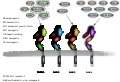
 Activation of ERBB kinases.png 678 × 604; 253 KB
-
-
-
-
-
-
-
-
-
-
-
-
-
-
-
-
 Binding of ATP to kinase active site of EGFR.svg 437 × 287; 106 KB
-
Characterizing-Tyrosine-Phosphorylation-Signaling-in-Lung-Cancer-Using-SH2-Profiling-pone.0013470.s012.ogv 1 min 9 s, 640 × 480; 1.46 MB
-
Coarse-Grained-Molecular-Simulation-of-Epidermal-Growth-Factor-Receptor-Protein-Tyrosine-Kinase-pcbi.1003435.s004.ogv 1 min 14 s, 512 × 512; 41.03 MB
-
Deep-and-high-resolution-three-dimensional-tracking-of-single-particles-using-nonlinear-and-ncomms8874-s2.ogv 36 s, 1,200 × 750; 12.68 MB
-
-
-
Deep-and-high-resolution-three-dimensional-tracking-of-single-particles-using-nonlinear-and-ncomms8874-s5.ogv 45 s, 1,024 × 768; 25.42 MB
-
Deep-and-high-resolution-three-dimensional-tracking-of-single-particles-using-nonlinear-and-ncomms8874-s6.ogv 45 s, 1,024 × 768; 19.12 MB
-
Deep-and-high-resolution-three-dimensional-tracking-of-single-particles-using-nonlinear-and-ncomms8874-s7.ogv 45 s, 1,024 × 768; 36.76 MB
-
 DL20230205 targeting-EGFR.tif 3,543 × 4,000; 5.86 MB
-
-
-
-
-
-
-
-
 EGF receptors and ligands.svg 976 × 670; 88 KB
-
 EGFR kinase (9125080896).jpg 661 × 747; 49 KB
-
 EGFR kinase covalently bound to HKI-272.png 978 × 688; 330 KB
-
 Egfr-erk pathway.png 2,396 × 2,488; 327 KB
-
 EGFR.jpg 599 × 703; 86 KB
-
-
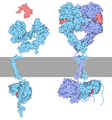
 Epidermal Growth Factor Receptor (EGFR) with Spitz protein.png 1,280 × 980; 500 KB
-
-
-
-
From-pathway-to-population-–-a-multiscale-model-of-juxtacrine-EGFR-MAPK-signalling-1752-0509-2-102-S3.ogv 43 s, 1,201 × 901; 22.59 MB
-
From-pathway-to-population-–-a-multiscale-model-of-juxtacrine-EGFR-MAPK-signalling-1752-0509-2-102-S4.ogv 43 s, 1,201 × 901; 32.97 MB
-
 Gefitinib 3d.png 902 × 714; 290 KB
-
-
-
-
-
-
 HPA EGFRcompilation DL20221120.tif 4,016 × 1,417; 4.67 MB
-
Human-neural-progenitors-express-functional-lysophospholipid-receptors-that-regulate-cell-growth-1471-2202-9-118-S1.ogv 2 min 1 s, 320 × 240; 6.06 MB
-
Human-neural-progenitors-express-functional-lysophospholipid-receptors-that-regulate-cell-growth-1471-2202-9-118-S2.ogv 1 min 14 s, 320 × 240; 3.45 MB
-
 Mutated EGFR kinase domain in complex with gefitinib.png 577 × 595; 297 KB
-
 Necitumumab EGFR 6B3S.png 2,048 × 2,048; 1.88 MB
-
-
-
New-Striatal-Neurons-in-a-Mouse-Model-of-Progressive-Striatal-Degeneration-Are-Generated-in-both-pone.0025088.s004.ogv 1 min 1 s, 1,348 × 939; 8.44 MB
-
 PBB GE EGFR 201983 s at fs.png 732 × 530; 11 KB
-
 PBB GE EGFR 201983 s at.png 255 × 135; 706 bytes
-
 PBB GE EGFR 201984 s at fs.png 732 × 530; 8 KB
-
 PBB GE EGFR 201984 s at.png 255 × 135; 450 bytes
-
 PBB GE EGFR 210984 x at fs.png 732 × 530; 11 KB
-
 PBB GE EGFR 210984 x at.png 255 × 135; 878 bytes
-
 PBB GE EGFR 211550 at fs.png 732 × 530; 12 KB
-
 PBB GE EGFR 211550 at.png 255 × 135; 926 bytes
-
 PBB GE EGFR 211607 x at fs.png 732 × 530; 12 KB
-
 PBB GE EGFR 211607 x at.png 255 × 135; 906 bytes
-
 Residues binding spitz protein and EGFR (left side).png 1,368 × 1,046; 943 KB
-
 Residues binding spitz protein and EGFR (right side).png 1,200 × 1,048; 910 KB
-
 Shc EGFR.svg 300 × 300; 108 KB
-
-
-
Specific-Visualization-of-Glioma-Cells-in-Living-Low-Grade-Tumor-Tissue-pone.0011323.s010.ogv 2.6 s, 524 × 512; 367 KB
-
Specific-Visualization-of-Glioma-Cells-in-Living-Low-Grade-Tumor-Tissue-pone.0011323.s011.ogv 2.6 s, 514 × 512; 357 KB
-
Specific-Visualization-of-Glioma-Cells-in-Living-Low-Grade-Tumor-Tissue-pone.0011323.s012.ogv 6.4 s, 516 × 512; 552 KB
-
Specific-Visualization-of-Glioma-Cells-in-Living-Low-Grade-Tumor-Tissue-pone.0011323.s013.ogv 3.2 s, 516 × 512; 341 KB
-
Specific-Visualization-of-Glioma-Cells-in-Living-Low-Grade-Tumor-Tissue-pone.0011323.s014.ogv 3.2 s, 530 × 512; 748 KB
-
-
-
-
 Varlitinib.png 286 × 144; 6 KB


.jpg)


_with_Spitz_protein.png)


.png)
.png)

